Bone Cross Section View / "Bone Cross Section" for Radius Digital Science on Behance / So without further ado, listen as annie narrate's her process of rendering out bone cross sections for medical illustration in a photoshop.
byAdmin-
0
Bone Cross Section View / "Bone Cross Section" for Radius Digital Science on Behance / So without further ado, listen as annie narrate's her process of rendering out bone cross sections for medical illustration in a photoshop.. (b) in this micrograph of the osteon, you can clearly see the concentric lamellae and central canals. • a local cross section uses a breakout to see through an. Hope you enjoy and please. There are trabeculae in spongy bone which gives its sponge like appearance. These pores serve to hold not only some marrow, but also nerves and vessels that transport blood to the cells delivering nourishment and gas exchange.
As the names suggest compact bone looks compact and the spongy bone looks like sponges. Click on the tags below to find other quizzes on the same subject. Hi all, i have uploaded a new medical animation tutorial. Find the perfect human bone cross section stock photo. Bone is found in the shafts of long bone and consists of various cylindrical units named as haversian system 47.
Cross Section of a Bone - Biology Forums Gallery from biology-forums.com From wikimedia commons, the free media repository. Select from premium bone cross section of the highest quality. This is a short tutorial using blender 2.8 that shows how to create a bone cross section and using images to create the textures. A cross section of a compact bone shows concentric circles called lamellae. (b) in this micrograph of the osteon, you can clearly see the concentric lamellae and central canals. Cross section through middle metacarpal bones of vector. Each system contains haversian canals surrounded by concentric lamellae of bone tissue 48. In this short video i use blender 2.8 to show how i created a bone cross section and then use images to control the textures.
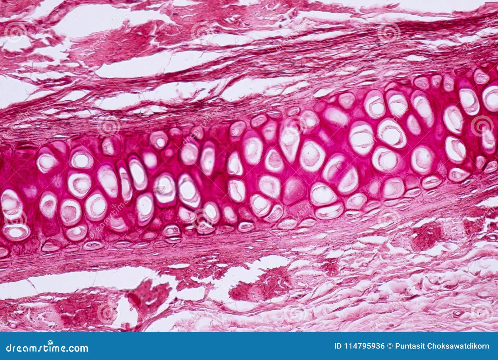
This is a cross section through decalcified bone.
They are obtained by taking imaginary slices perpendicular to the main axis of organs, vessels, nerves, bones, soft tissue, or even the entire human body. Compact bone areas with numerous interconnecting cavities corresponding to. I don't find it enhances the image. • a half cross section shows a cross section of the model on one side of a selected plane, but not on the other side, as shown below. I am not an expert on this subject, so i was wondering if anyone could put their input on this image. Bone decalcification is the removal of the mineral component using an acid, leaving the bone soft and easy to cut. This is a cross section through decalcified bone. A cross section of a compact bone shows concentric circles called lamellae. These pores serve to hold not only some marrow, but also nerves and vessels that transport blood to the cells delivering this simply involves placing a section of the bone on the microscope stage and viewing. Cross section through middle metacarpal bones of vector. Viewing leaf structure under the microscope shows different types of cells that serve various functions. Bone cross section — stock image. There are two ways to study bone histology.
Why is the marrow red? They are obtained by taking imaginary slices perpendicular to the main axis of organs, vessels, nerves, bones, soft tissue, or even the entire human body. I am not an expert on this subject, so i was wondering if anyone could put their input on this image. (b) in this micrograph of the osteon, you can clearly see the concentric lamellae and central canals. Hope you enjoy and please.
Cross Section Human Cartilage Bone Under Microscope View ... from thumbs.dreamstime.com As shown in figure 2. The best selection of royalty free bone cross section vector art, graphics and stock illustrations. Why is the marrow red? This is a cross section through decalcified bone. Dreamstime is the world`s largest stock photography community. • a full cross section displays the cross section across the whole view, as shown in the two examples above. In dry ground bone, the organic. In a cross section of a bone you can usually see two types of bone tissue what are these called?
Start studying bone cross section.
In this short video i use blender 2.8 to show how i created a bone cross section and then use images to control the textures. Each system contains haversian canals surrounded by concentric lamellae of bone tissue 48. • a local cross section uses a breakout to see through an. In a cross section of a bone you can usually see two types of bone tissue what are these called? Don't assume that the cross sectional area is the same no matter where you cut. Use them in commercial designs under lifetime. These pores serve to hold not only some marrow, but also nerves and vessels that transport blood to the cells delivering this simply involves placing a section of the bone on the microscope stage and viewing. Start studying bone cross section. Bone cross section — stock image. Find the perfect bone cross section stock photos and editorial news pictures from getty images. This is a cross section through decalcified bone. Department of histology, jagiellonian university medical under the stereo microscope (and tem images of bone that was sectioned perpendicular to the collagen fibril axes appear strikingly different from longitudinal views (figure 6). Bone cross section — stock image.
In a cross section of a bone you can usually see two types of bone tissue what are these called? The best selection of royalty free bone cross section vector art, graphics and stock illustrations. A cross section of a human long bone. From wikimedia commons, the free media repository. There are trabeculae in spongy bone which gives its sponge like appearance.
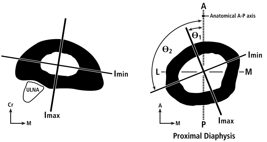
Biomechanics of Bone | Team Bone from teambone.com Don't assume that the cross sectional area is the same no matter where you cut. Huge collection, amazing choice, 100+ million high quality, affordable rf and rm images. Each system contains haversian canals surrounded by concentric lamellae of bone tissue 48. Your bone cross section stock images are ready. If your post declares something as fact, please cite a source in it, or in the comment section. • a full cross section displays the cross section across the whole view, as shown in the two examples above. Use them in commercial designs under lifetime. Dreamstime is the world`s largest stock photography community.
As the names suggest compact bone looks compact and the spongy bone looks like sponges.
They are obtained by taking imaginary slices perpendicular to the main axis of organs, vessels, nerves, bones, soft tissue, or even the entire human body. There are two ways to study bone histology. Select from premium bone cross section of the highest quality. There are trabeculae in spongy bone which gives its sponge like appearance. • a local cross section uses a breakout to see through an. Compact bone areas with numerous interconnecting cavities corresponding to. This is a cross section through decalcified bone. Hi all, i have uploaded a new medical animation tutorial. A cross section of a human long bone. Find the perfect bone cross section stock photos and editorial news pictures from getty images. Don't assume that the cross sectional area is the same no matter where you cut. (b) in this micrograph of the osteon, you can clearly see the concentric lamellae and central canals. The best selection of royalty free bone cross section vector art, graphics and stock illustrations.
We are sharing her tutorials on street anatomy over a series of posts to help get these out to as many artists as possible! bone cross section. This is a short tutorial using blender 2.8 that shows how to create a bone cross section and using images to create the textures.